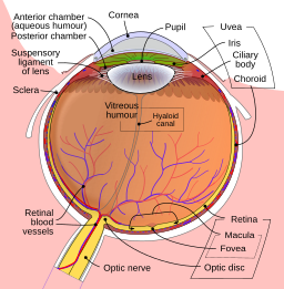
Glaucoma is a group of diseases that cause damage to the eye’s optic nerve and can ultimately result in loss of vision and blindness. Extensive research has indicated that eye pressure is an important element when it comes to glaucoma. It is a major risk factor for optic nerve damage. Glaucoma has been dubbed the silent thief of sight.
Process

The front of the eye has a space which is called the anterior chamber. A transparent fluid travels constantly in and out of the chamber and serves as nourishment for tissues that are in close proximity.
The fluid departs the chamber at an open angle where the cornea and iris meet. Once the fluid reaches this angle, it flows through a spongy meshwork and leaves the eye.
Glaucoma occurs when this fluid passes lethargically via the drain. Eventually, there is an assembly of fluid in the passage, which means it exerts pressure inside the eye up to a point where it can impair the optic nerve. When damage is inflicted on the nerve, glaucoma, and loss of vision abound. This is precisely why governing the pressure inside the eye is so critical.
Types
The broad categories of glaucoma are open-angle and narrow-angle glaucoma. The ‘angle’ is a reference to the drainage angle inside the eye which controls the discharge of fluids (aqueous), which are continually produced in the eye.
If the aqueous reaches the drainage angle, the glaucoma is known as open-angle. If for some reason the angle is not accessible, then that is classified as narrow-angle glaucoma. Now let’s discuss the various subtypes.
Primary Open Angle Glaucoma
This is a common occurrence that steadily lowers the person’s capability of peripheral vision without the exhibition of other symptoms. A concern for those affected is that by the time they observe it or their doctor is able to detect it, significant and irreparable damage may already be caused.
If the patient’s intraocular pressure remains high, the havoc wreaked by primary open-angle glaucoma may carry on until tunnel vision is developed. This means there may be a point where the patient is only able to see people or objects that lie straight ahead and not sideways. The concern of eventuality is that blindness may transpire as well.
Normal-Tension Glaucoma
Also called normal pressure or low tension glaucoma, it is an open-angle affliction. It instigates visual field loss since the optic nerve is at a detriment. However, patients who have normal-tension glaucoma do not experience a decline in their intraocular pressure levels.
Furthermore, this form of glaucoma is innocuous, and permanent harm may not be noticed until symptoms like tunnel vision occur. Although many doctors believe it is linked to inefficient blood flow to the optic nerve, the exact cause of normal-tension glaucoma has not been determined yet.
Pigmentary Glaucoma
This is a rare form of glaucoma that is the outcome of congestion of the drainage angle of the eye courtesy of pigments that have severed from the iris. This reduces the aqueous outflow from the eye over a period of time. Naturally, an inflammatory response to the blockage causes disturbance to the drainage mechanism.
Secondary Glaucoma
If the patient shows symptoms of chronic glaucoma in the aftermath of an eye injury, then that could be a sign that they are suffering from secondary glaucoma. This may also develop if an eye infection is active or if there is discernible inflammation. A cataract may result in an enlargement of the lens, which could end up causing secondary glaucoma as well.
Congenital Glaucoma
Congenital glaucoma is an inherited form which means it may make its presence felt at birth. There is empirical evidence to suggest so since 80% of cases are diagnosed by the time a child is one year old. These children have a malfunctioning drainage system in their eyes.
Moreover, it can be hard to observe signals of congenital glaucoma, since children are too young to comprehend the illness and voice their own opinion regarding the matter. This is where the parents play a decisive role. If they notice that their child’s eye appears hazy or enlarged, they should consult a doctor at the first instance.
Acute Angle Closure Glaucoma
Finally, this is a form of narrow-angle glaucoma. This ailment produces symptoms abruptly which include pain in the eye, headaches, halos around lighting, dilated pupils, and nausea. All of the above constitute a medical emergency. Acute angle glaucoma may last for a few hours at a stretch and then subside. It may reappear for another bout. In extreme cases, it may last long without even momentary relief.
Treatments

If treatment is availed promptly, at the very least it can stem the progression of the disease. This is one of the reasons patients should see a doctor as soon as possible. Of course, medication is a viable solution for rectifying glaucoma. These usually come in eye drops or pills. Also, they are generally administered early on before other avenues are explored. They can lower eye pressure and hinder the production of excess fluids.
Most medicines taken to cure glaucoma must be taken consistently as directed by the professional. There are some that introduce side effects, such as a stinging sensation or recognizable redness in the eyes.
Laser trabeculoplasty is a procedure that aids the eyes in draining out abundant fluids. The operation is performed at the doctor’s clinic. It begins with the application of numbing drops to the affected eye or eyes. Once the patient is seated and facing the laser machine, the doctor exposes them to a special lens. A high-density beam is aimed through the lens and deflects onto the meshwork inside the eyes. The laser creates space for the fluids to drain more efficiently. There are potential side effects of laser trabeculoplasty, one of which is inflammation. Medication may still be required post-surgery.
Another feasible resort is conventional surgery. This procedure creates a new passage for the fluids to be discharged. Doctors may suggest this treatment if laser surgery is not considered effective, depending on the case at hand.
Also called trabeculectomy, it is performed in an operating room. Medication is administered pre-surgery to assuage the patient. Small injections are made around the affected eye to numb it. A small tissue is removed to make a new avenue for the fluid to flow.
Once the surgery is complete, eye drops must be taken for weeks. In most cases, the operation is only carried out on one eye. If the other eye is affected, then another surgery will be scheduled a few weeks later. Conventional surgery is effective in roughly 70% of all cases.
Make sure you make responsible visits to your optometrist. Like all other types of routine doctor visits, don’t lose sight of your eye’s health (pun intended!).
