What Causes Back Pain?

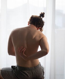
Man putting his hands on his back
Photo by Adrian “Rosco” Stef on Unsplash

If you’re reading this, you or someone you care about has likely experienced back pain. According to goodbody.com, as many as 540 million people suffer from one type of back pain or another.

It is one of the most common reasons for doctor visits and missed workdays. Fortunately, there are a variety of ways to reduce your risk of experiencing back pain. This blog post will explore the various causes of back pain. If you’d like to know some preventative techniques and management of back pain, please check out our article on What to Do About Back Pain.

What is the Most Common Cause of Back Pain?

Our muscles rely on a network of nerves that travel through them to deliver signals to the brain. When these nerves are overstretched, as can happen with muscle strains, they can get pinched and cause pain.

Disc problems, technically called intervertebral discs, are another common cause of back pain. The discs that cushion the vertebra in the spine are prone to wear and tear as we age, which increases the risk of the disc rupturing.

They is made up of a gel-like substance, and if the pressure within the disc becomes too great, it will rupture and the gel will leak out. The disc can then press against the surrounding nerves, which will then send pain signals to your brain.

These discs located in the lumbar region of the lower spine are particularly prone to herniation, otherwise known as herniated discs. This is not only the most common cause of back pain in this part of the spine, but it is also the leading cause of sciatica (see below).

Disc Problems

Illustration of spine/disc degeneration
Disc Degeneration: Photo: IStock

Intervertebral discs are soft, spongy pads made of a gelatinous substance called a disc nucleus. It lies between each of the vertebrae to provide a cushion and to allow the vertebrae to rotate.

If the disc nucleus becomes too thick or ruptures, the disc may be unable to cushion the vertebrae properly. It may press against the surrounding nerves or cause irritation that travels down the legs as sciatica.

These kinds of disc problems can lead to degenerative disc disease – when the disc becomes thinner and less able to cushion the vertebrae. This can occur during the normal aging process when the disc may begin to break down.

What Causes Disc Denegation?

      • Age: The softcore that we mentioned contains mostly water. As you age, it can lose some water, causing discomfort.
      • Injuries: No doubt any injury can lead to a problem and that includes injuries in your spine.

Poor Posture

Woman slouching over on a chair looking at a laptop
Photo by Christina @ wocintechchat.com on Unsplash

Sitting is the new cancer! Have you heard that before? Poor posture can occur when you spend long hours sitting or slouching or have an overly stiff spine.

When the spine is in a neutral position, it is straight and flexible. However, when you spend hours at a time in a hunched position or with a stiff spine, the muscles and ligaments supporting the spine become shortened.

Over time, these shortened muscles and structures can put excessive pressure on the spine. The spine is composed of bones that are held together by ligaments, muscles, and other soft tissues. These structures may not be able to withstand this pressure. Squeezing the spinal discs may cause the disc fluid to leak out, which may irritate nearby nerves and cause pain.

The Types of Back Pain that Are a Cause for Concern

Certain types of back pain can indicate a serious underlying medical condition. The first step in determining whether or not your back pain is cause for concern is to determine what type of pain you are experiencing.

    • Herniated Discs: A herniated disc occurs when the soft, spongy center of a disc pokes through the disc wall and presses against the surrounding nerve roots. It is one of the most common causes of sciatica and back pain.
    • Sciatica: Sciatica is pain that travels down one or both legs. It is often caused by the irritation of one or more sciatic nerve roots due to a herniated disc.
    • Lumbar Spinal Stenosis (LSS): Lumbar spinal stenosis is a condition in which the spinal canal narrows. This can put pressure on the spinal cord and nerves, which can cause back pain. This is one of the most common causes of back pain that is a cause for concern.

Conclusion

Back pain is a common ailment that can affect anyone at any stage in their life. Luckily, there are lots of ways to prevent and manage back pain.

The most important thing you can do is stay active by following a healthy diet plan and engaging in daily exercise. Stay hydrated, and be sure to get enough rest so that your body can heal properly when it does sustain an injury.

If you do experience back pain, see your doctor as soon as possible. It is often an indication of a more serious underlying condition, such as a possible disc problem.

Your doctor can perform diagnostic tests and recommend treatment options that can help you avoid further complications down the road.

The Bones in Our Hands

In continuation of our series about our bone structure, we will now discuss our hands. Probably of all the bones in our bodies, the hands have the most flexible bone assembly and for good reason. Let’s take a look at how these bones and ligaments work to allow us this extreme flexibility of movement.

OK, so what are the bones in our hands?

There are 14 bones in each finger (and toes too!). Of these, they are broken down into three bone groups: Phalanges, Metacarpal and Carpal.

Phalanges

These are the actual fingers of your bones. They extend from the very tip of your nails down to the part when you can bend your fingers. The phalanges are then broken down into three additional categories: the distal, middle and proximal, with the exception of the thumb which has only two areas.

      • The distal phalanges are your fingertips. Ever hear the expression “sensitive to the touch”?, Well, it is these bones that are responsible for that! They have nerve endings that are designed to interpret the feelings of touch on your fingers into nerve impulses that are channeled to the brain.
      • The middle phalanx is just that, the middle bone section of the phalange It is connected to the distal phalanx at the top and the proximal phalanx at the bottom.
      • The proximal phalanx is the largest of the three bones in the finger, joined by the metacarpal and the middle phalanx as shown in the diagram.

Metacarpal bones

There is one of these bones with each finger, labeled Metacarpal I – Thumb, Metacarpal II – Index finger, Metacarpal III – Middle finger. Metacarpal IV – Ring finger and Metacarpal V – Little finger.

Carpal bones

The bones are located in the wrist area. There are eight of them and are relatively small compared to the other bones in the hands. The carpal are the bones that connect the hand bones to the forearm.
Heard of carpal tunnel? This happens when the nerves, namely the median nerve receives too much pressure, resulting in certain pain and if not resolved, can lead to nerve damage.

The median nerve is one of a group of nerves that originate in the neck. They combine to form a single nerve through the arm. The nerve then continues down the arm to the wrist and then into the hand.

Symptoms

      • Tingling and numbness in your fingers or hand. Usually the thumb and index, middle or ring fingers are affected, but not your little finger.
      • Weakness is another symptom.

Treatment

Carpal tunnel syndrome does not require hospitalization, rather standard home procedures should cure it. Some of treatments you can try are:

      • Take breaks from repetitive tasks, such as typing, which is the most popular method of acquiring the pain.
      • Stretch your hands and wrists whenever possible.
      • Try over-the-counter (OTC) medications.

Other Hand Issues

Arthritis

Arthritis, a common disorder that affects about 54 million people, almost all adults are diagnosed with arthritis. It refers to an inflammation of one or more of the joints. There are two types of arthritis that affect the joints of the hand and wrist. Osteoarthritis and rheumatoid arthritis.

Osteoarthritis. AKA wear-and-tear arthritis mainly affects seniors and is caused by the cartilage in joints that wear away over time.

Rheumatoid arthritis is the result of the lining of the joints to swell.

Both can be painful, but over the counter medications can be the treatment for most people. If meds do not work, your doctor can decide the next step up to surgery, but usually meds will do the trick.

Conclusion

Our hands have evolved over millions of years to be the useful tools they are today. We share these gifts of nature with thousands of other species on the planet, but only now are we moving into a new generation where human hands will not be human, but android. Let’s hope they will be put into as good use as they are with we humanoids.

The Human Ribs

We at Howard Fensterman Charities believe that the more you know about your health, the better you are to identify ailments that may affect you during your lifespan or perhaps you just want to learn more about you your body functions. Either way, we welcome you here and hope you gain the knowledge you are seeking.

Drawing of the human ribsOur Ribs

Before we delve into our rib discussion, let’s lay the groundwork regarding some terms we should be familiar with.

Terms Associated with the Human Rib Cage

Rib Cage: So, we start with the rib cage, also known as the thoracic cage – what is it? Quite simply, it is the ribs that are attached along the vertebral column (spine).

Thoracic Vertebrae: These are the bones along the vertebral column between the cervical bones above and the lumbar vertebrae.

Sternum: Also known as the breastbone, it is a long flat bone situated in the central part of the chest. It connects to the ribs and forms the front of the rib cage and in doing so, it encloses and protects the heart and lungs from injury. It is one of the largest flat bones of the body.

Human Ribs Explained

X-Ray view of human ribs
X-Ray view of the rib cage

The human species have 24 ribs, separated in 12 pairs and are attached at the back to the thoracic vertebrae (spine). They are numbered from 1–12 (more on this later). Essentially, they are the bones that curve around the spine and form the majority of the thoracic cage. They are lightweight, but strong and are designed to protect the heart and lungs, also known as the thoracic organs.

There are 12 ribs in all on each side of the vertebral column and are designated into three categories: true (vertebrosternal) ribs, false (vertebrochondral) ribs and floating (vertebral, free) ribs. True ribs are the ones that attach directly to the sternum and are numbered one to seven. False ribs are numbered eight to ten are indirectly connected to the sternum.

What Happens When You Break a Rib?

Broken ribs are one of the most common injuries when it comes to fractured bones. The injuries are usually associated with sports or vehicle accidents. Generally speaking, broken ribs aren’t dangerous, but if they are cracked into numerous pieces, that could pose a problem. Similar to broken glass, there may be sharp or jagged edges stemming from one of the fractures and that can cause damage to internal organs or major blood vessels.

With that said, if you incur a broken rib, see a doctor. In most cases, fractured ribs can heal on their own in a few months, but it goes without saying that anytime you fracture a bone, you should seek medical help to be sure your health is not in jeopardy.

What are the Symptoms of Broken Ribs?

You may feel chest pressure or squeezing pain. If you do feel this, don’t second guess it because it may also be associated with a heart attack.

How Can I Prevent a Broken Rib?

Essentially you can’t if it occurs from an injury, but you can do some preventative maintenance such as:

    • Wear the appropriate protective equipment in sports.

    • Keep your floors clear of debris. One fall and you can get a bone broken. This is especially important for the elderly. Place a rubber mat in the shower.

    • Eat the right foods that contain calcium and vitamin D which is essential for maintaining strong bones. 1,200 milligrams of calcium and 600 International Units (IU) of vitamin D daily from food and vitamin supplements are recommended.

What is the Human Spine?

In our previous article, we discussed an overview of our bones and what they do (besides the obvious). Now let’s delve a bit deeper to learn what each section of our skeleton does, starting with the spine.

What is the Structure of the Spine?

Human Spine
Photo: Wikimedia (public domain)

The spine, also known as the vertebral column is that long curvey bone that we see on posters when we’re in the doctor’s office. It supports our upper body weight. It runs from the base of the skull to the tailbone, also known as the coccyx.

The spine supports our posture but allows us to move while allowing for movement. It also protects the spinal cord.

This column contains 26 bones in adults which include 24 separate vertebrae (interlocking bones that form the spine), but children (adolescent and younger) have 33 bones in the spine because the sacrum (a triangular set of five bones at the base of the spine just above the coccyx) does not fuse with the coccyx until adulthood.

Child-Adult Spine Increase
Child Spine Increases After Adulthood (Wikipedia Public Domain)

Vertebrae Structure

The vertebrae are divided up into three regions: cervical, thoracic and lumbar and contain several important parts: the body, vertebral foramen, spinous process and transverse process.

There are Five Major Regions of the Spine

    • Cervical: These are the vertebrae in the neck and form the cervical region of the spine. There are seven of them. They are the thinnest vertebrae in the spine, but they provide great flexibility to the neck area.
    • Thoracic: There are 12 vertebrae connected here and are in the chest area, known as the thoracic region. They are larger and stronger than the cervical vertebrae but are not as less flexible as the ones in the neck region.
    • Lumbar: You might have heard on commercials about the lumbar. These are five vertebrae in the lower back because there are many people who have issues in this area due to the fact that all of the upper body’s weight puts pressure on this area. These vendors offer cushions that help strengthen the lumbar area. Lumbar vertebrae are stronger than the thoracic vertebrae, but not strong enough to cause some people periodic pain.
    • Sacral: The sacrum is a single bone in the adult skeleton that is formed by the fusion of the five smaller vertebrae when adulthood starts.
    • Coccygeal: As the name suggests, this is where the coccyx resides. It is a single bone in the adult skeleton and is formed by the fusion of four vertebrae at the beginning of adulthood. Also known as the tailbone, it holds our body weight when we sit.

The Spine Numbering System

The Human Spine
Photo: Wikipedia

The spine is numbered by its the first letter of their vertebrae regions; accordingly, there are five categories of designation.

At the top of the vertical column are the seven cervical spinal nerves labeled C1 through C8. Then there are the 12 thoracic nerves, which are labeled T1 through T12.

Similarly, there are the 5 lumbar spinal nerves titled L1 through L5 and under the lumbar are the 5 sacral spinal nerves S1 through S5 and then the coccygeal of which there is 1.

What Are Some Diseases Associated with the Spine?

Otherwise known as back pain, there are many illnesses associated with the spine. There is Kyphosis, an abnormally excessive curvature of the spine and happens when the vertebrae in the upper back area become more wedge-shaped.

There also herniated disks. Known as the vertebral discs they have the function of shock absorption. With this disc becomes ruptured, it becomes smaller and is no longer able to provide as the amount of shock absorption is was initially intended for. This can result in back pain, sciatica and other disorders. Herniated discs can be caused by injury or from the normal wear and tear of aging. Treatment can be by medication and/or physical therapy and if these methods do not work, then surgery might be needed.

For a comprehensive list of spine disorders, please click here.


No Bones About It

Ever hear grandma say “Oh my aching bones”? What is she actually referring to? In order to answer that, we need to gather some preliminary information about our skeletal system and how it works.

Your Bones are Alive!

Human Bones in Vertical Position
Photo by camilo jimenez on Unsplash

Don’t think for one second that just because they are hard like a rock, you might think that they perform no service to the body, contrary to our organs like the heart and lungs which maintain blood flow and help us healthy.

What Are Bones Made of?

Your bones are made up of virtually the same materials. Here are the layers that comprise the bones:

      • periosteum – dense membrane that contains the nerves and blood vessels.
      • compact bone. This layer is smooth hard. It’s the part you see when you look at a skeleton.
      • cancellous – looks a bit like a sponge, but it’s actually very strong.
      • bone marrow looks like jelly and its job is to make blood cells.   

Here’s a brief overview of the bone sections in your body:

The Spine

The spine is the one part that gives you that flexible ability to twist around. It is actually a combination of 33 bones. They are referred to as vertebrae and each of these 33 vertebrae formed like a ring. Put your hands on your back and you can actually feel it! (Wait for our upcoming article that details the vertebrae.)

The Ribs

Human Skeletons
Photo by Danielle MacInnes on Unsplash

Think of your ribs as the security guard that circles your heart and lungs. The ribs are broken up in twos –  left and right sides and each side is identical to the other. 12 sets of ribs are what most people have. Every one of the 12 sets of ribs appends to the back to the spine, where they are held by the thoracic vertebrae. The initial seven sets of ribs append in the front to the sternum – a solid bone in the focal point of your chest that holds those ribs together.

Your skull protects the most important part of all, the brain. You can feel your skull by pushing on your head, especially in the back a few inches above your neck. The skull is actually made up of different bones. Some of these bones protect your brain, whereas others make up the structure of your face. If you touch beneath your eyes, you can feel the ridge of the bone that forms the hole where your eye sits. And although you can’t see it, the smallest bone in your whole body is in your head, too.

The stirrup bone behind your eardrum is only .1 to .13 inches long. Your lower jawbone is the only bone in your head that can move. It opens and closes to let you talk and chew. 

The Hands

We’ve all heard of carpal tunnel. Well, that is one factor that can happen to your hands, As you sit and type at the keyboard, even when you pick up a phone, you’re using the bones in your fingers, hand, wrist, and arm. Each arm is attached to a shoulder blade or scapula, a large triangular bone on the upper back corner of each side of the ribcage.

The arm is made up of three bones: the humerus which is above your elbow, and the radius and ulna,  which are below the elbow. The center part of your hand is made up of five separate bones.

The Legs

Your legs are attached to a circular group of bones called your pelvis. The pelvis is a bowl-shaped structure that supports the spine. It is made up of the two large hip bones in front, and behind are the sacrum and the coccyx. The pelvis acts as a tough ring of protection around parts of the digestive system, parts of the urinary system, and parts of the reproductive system.

Leg bones are very large and strong to help support the weight of your body. The bone that goes from your pelvis to your knee is called the femur and it’s the longest bone in your body. 

The Joints

The place where two bones meet in your body is called a joint. Some joints move and others don’t. Fixed joints are fixed in place and don’t move at all. Your skull has some of these joints which close up the bones of the skull in a young person’s head.

Moving joints are the ones that let you twist and turn. Other joints move a lot. One of the main types of moving joints is called a hinge joint. Your elbows and knees each have hinge joints, adequately called that because they are similar to hinges on a door.

Similar to putting oil on a hinge to make it stop squeaking? The joints come with their own special fluid called synovial fluid that helps them move freely. Bones are held together at the joints by ligaments which are like very strong rubber bands.

Conclusion

All in all, your bones play a significant role every day. Stay tuned for a detailed account of just how each of these bone sections works!

Spinal Cord Injuries – Types, Levels and Implications

 Image of spine with all 33 vertebras
Human spine (Photo by Pixaby)

Spinal Cord Overview 

“Oh my aching back!”. A term uses so much by so many, it becomes almost status quo. But what causes one to feel this pain? Let’s take a look.

The spinal cord is the most integral neural pathway in the human body. It connects the brain with the nervous system which serves as a relay between the brain, spinal cord and rest of the body. 

Since it is the central neural pathway, the spinal cord is a collection of nerves. In fact, there are 31 pairs of nerves which originate from the spinal cord and travel to different parts of the body. Through these nerves, the brain sends commands to different muscles and organs and controls their movement. There is another set of nerves which travels from different organs and connect to the spinal cord. These nerves are responsible for bringing in information from the organs to the brain. Examples of such nerves include the nerves travelling from the hands and legs which bring in sensory information. 

The spinal cord is protected through a bony column called the spine. The spine is divided into 33 vertebrae which are individual pieces of bones locked together. The vertebrae are divided into five regions namely cervical (7), thoracic (12), lumbar (5), sacrum (5), and coccyx (3-5). 

Spinal Cord Injuries 

The spinal cord is one of the most sensitive parts of the human body. This is because, once damaged, the spinal cord cannot repair itself. The spinal cord injury can result from a physical trauma such as an accident, fall or slip. It can also be a result of compression of the spinal cord due to the growth of tumor in the body. Another possibility which can result in a spinal cord injury is the loss of normal blood supply to the spinal cord. 

In the US, every year, approximately 1,200 new cases of spinal cord injury are reported every year. The condition is more common among young, white males.  

There are two types of spinal cord injuries.

    • Complete spinal cord injury 
    • Incomplete spinal cord injury 

 Let’s take a look at each one.  

Complete Spinal Cord Injuries 

In case of complete spinal cord injury, there is a complete or total loss of sensation and bodily functions in the area below the site of injury. This means that if there is a complete spinal cord injury in the lumbar region of the spine, all muscular functions below that region will be compromised including bowel movement and function of legs. Such a person will not have any sensation or control over his body below the site of injury.   

Incomplete Spinal Cord Injuries

In case of an incomplete spinal cord injury, a person can feel some sensation and there is remaining function in and below the affected region. For example, if the thoracic region is injured, in case of incomplete spinal cord injury, the person will still feel the sensation of pain and pressure and there will be muscular function in the region below the thoracic cavity. 

Regardless of the type of spinal cord injury, both sides of the body are likely to get affected.   

Levels and Implications of Spinal Cord Injury

The implications of spinal cord injury significantly depend upon the type and level of spinal cord injury. As mentioned earlier, the spine is divided into five regions, and the level of spinal cord injury is determined by the region of the spine affected by the injury. Below you will find out about the implications of spinal cord injury depending upon the level. 

Cervical Spinal Cord Injury 

The cervical region includes the first 7 vertebrae in the neck region. Since it is closest to the brain, spinal cord injuries in the cervical region are most dangerous. 

Implications of Cervical Spinal Cord Injury 

This level of spinal cord injury is associated with complete or partial loss of sensory or muscular function in the body. Most often, in case of a cervical spinal cord injury, the patient experiences tetraplegia or quadriplegia. This means that all four limbs of the person can get affected to some extent in case of an 

The injury of the upper vertebrae of this section is often more dangerous and can result in permanent damage to all four limbs. In case of a more severe injury, this level of spinal cord injury can prove to be fatal. But as you go down the cervical vertebrae, the risk decreases and often the injury is incomplete. 

Thoracic Spinal Cord Injury 

The thoracic spine includes 12 vertebrae which are numbered as T-1 to T-12. This section of the spine is basically the upper, and middle part of the back. The main function of this section of the spine is to ensure that you stay upright and maintain the right posture while sitting and standing. The nerves passing through these vertebrae control the movement of muscles in the upper chest including the muscles of the rib cage and diaphragm. They also include abdominal muscles and muscles of the upper back. 

Implications of Thoracic Spinal Cord Injury 

Injuries in the thoracic section of the spinal cord generally affect the lower part of the body. In the case of a thoracic spinal cord injury, the lower limbs of the person get affected. The bowel and bladder control is also compromised but usually, the arms and hands continue to function normally. 

Lumbar Spinal Cord Injuries 

The lumbar portion is the lowest and most important part of the spinal cord. It comprises five vertebrae. The vertebrae of this region are larger than the vertebrae of all other regions of the spine therefore this section carries the most weight of the spine. 

Implications of Lumbar Spinal Cord Injuries  

In the case of lumbar spine injuries, a person may experience complete or partial loss of function in the hips, legs, bladder and bowel control. It can also affect functions such as bending of the knees and foot. 

Sacral Spinal Cord Injuries 

This section of the spine is right above the tailbone which is known as the coccyx. The nerves of this region affect the function of the hips, pelvic organs, and reproductive organs. 

Implications of Sacral Spinal Cord Injuries 

Injuries in this section of the spinal cord result in loss of function in the hips and organs of the pelvic region including bladder and reproductive organs. 

Conclusion 

A spinal cord injury is dangerous because it can disrupt the communication process between the brain and the rest of the body. Physical trauma, compression due to a tumor, or loss of blood supply to the spinal cord can all lead to spinal cord injuries. There can be complete or incomplete spinal cord injuries which are based on the extent to which motor and sensory reflexes of a person are affected. Regardless of the type, if you look at the general implications of spinal cord injury, you will notice some of the following effects. 

    • Loss or reduced mobility, 
    • Changes in sensation, 
    • Modified sexual function,
    • Loss of bladder or bowel control and or,  
    • Intensified reflex actions or increased frequency of spasms.